A Quiet Transformation: The Rise of Ultrasound-Guided Regional Anesthesia
By Hajra Shereen, BS AHS (Anesthesia Technology), Department of MLT, Haripur, KPK, Pakistan.
Supervised by: Samsaam Fazal, MS-AHS
Affiliation: The University of Haripur
A quiet transformation is reshaping operating rooms, the rise of ultrasound-guided regional anesthesia. What was once a “blind” procedure relying on touch and anatomical landmarks has evolved into a precise, image-guided technique that enhances both patient safety and clinical confidence.
Regional anesthesia numbs a specific area of the body instead of inducing full unconsciousness. It includes spinal, epidural, and peripheral nerve blocks that allow patients to stay awake yet pain-free during surgery. In the past, anesthetists located nerves through experience and estimation, which sometimes caused incomplete blocks or accidental injury.
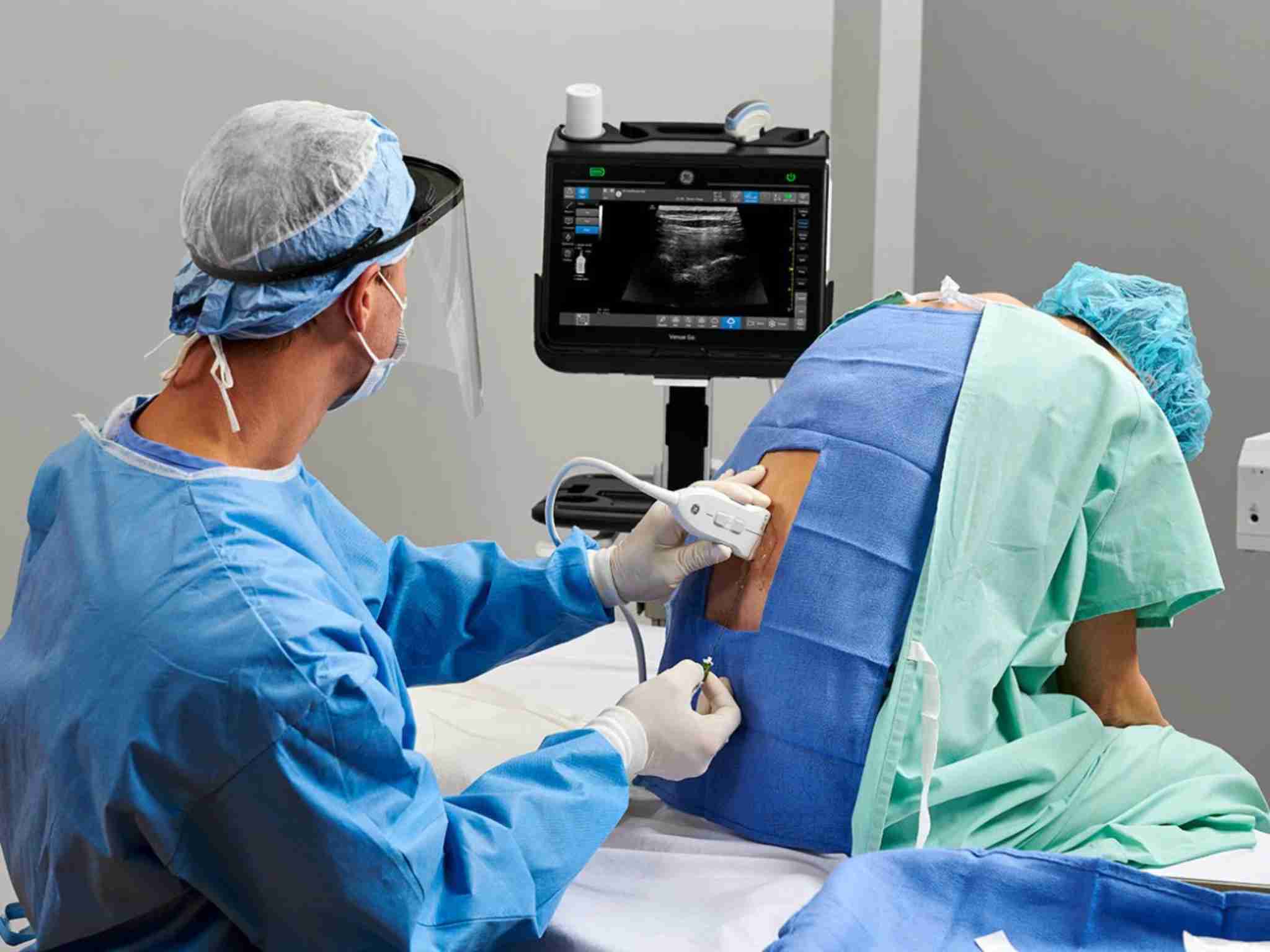
Today, ultrasound guidance provides real-time visualization of nerves, blood vessels, and the advancing needle tip. This accuracy minimizes complications and ensures the local anesthetic is delivered exactly where it is needed. It also allows safe anesthesia in patients with complex or unclear anatomy.
Beyond the operating room, ultrasound-guided regional anesthesia reduces the need for opioids, helping patients recover faster with fewer side effects such as nausea and drowsiness. Many hospitals now include it in Enhanced Recovery After Surgery (ERAS) programs, improving patient comfort and satisfaction.
For anesthesia technologists and practitioners, this advancement demands new skills, understanding sonoanatomy, using ultrasound machines, and maintaining sterile precision. The outcome is rewarding: safer procedures, faster recovery, and more confident teams.
Ultrasound has truly become the “stethoscope” of modern anesthesia turning invisible anatomy into visible safety and marking a major step toward patient-centered care.